Understand skin cancer surgery, the importance of biopsy and margins, and care after removal. Information to help you prepare for assessment and participate in decisions.
Has a lesion changed, bled or failed to heal?
Seek assessment. If you already have a confirmed diagnosis, mention it when booking and bring your pathology report. Understand urgency ↓
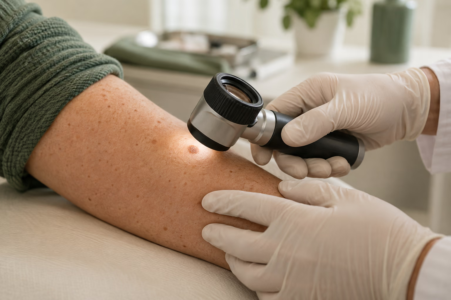
Illustrative dermoscopy scene showing assessment of an arm lesion, generated with AI. It does not depict a real consultation or patient.
Care begins with diagnosis.
01 / SKIN CANCER GUIDE
When should a skin lesion be assessed?
A sore that does not heal, spontaneous bleeding or a changing mole deserves assessment. These findings do not prove cancer, but should not be treated as a purely cosmetic concern.
Prompt appointment
Suspected or confirmed cancer
Tell the clinic if a pathology report has already confirmed skin cancer. Growth, changes in colour or shape, and a lesion returning at a previous surgical site warrant prompt review. Bring the report and earlier records.
Melanoma awareness
A mole unlike your others
Asymmetry, irregular borders, different colours and evolution are warning signs. Diameter can help with observation, but melanoma may also be small. Do not wait for every warning sign to appear.
Urgent medical care
Heavy bleeding or significant deterioration
Bleeding that will not stop, fever with a worsening wound or feeling seriously unwell needs same-day assessment. An emergency should not wait for a social-media reply.
Priority and surgical timing depend on tumour type, examination, pathology and general health. Not every suspicious lesion requires same-day surgery; indefinitely delaying investigation is also unsafe.[1][2][3]
02 / SKIN CANCER GUIDE
Three diagnoses, different decisions
Skin cancer includes diseases with different behaviours. Identifying the correct type is the first step in selecting treatment, deciding which tests are needed and planning follow-up.
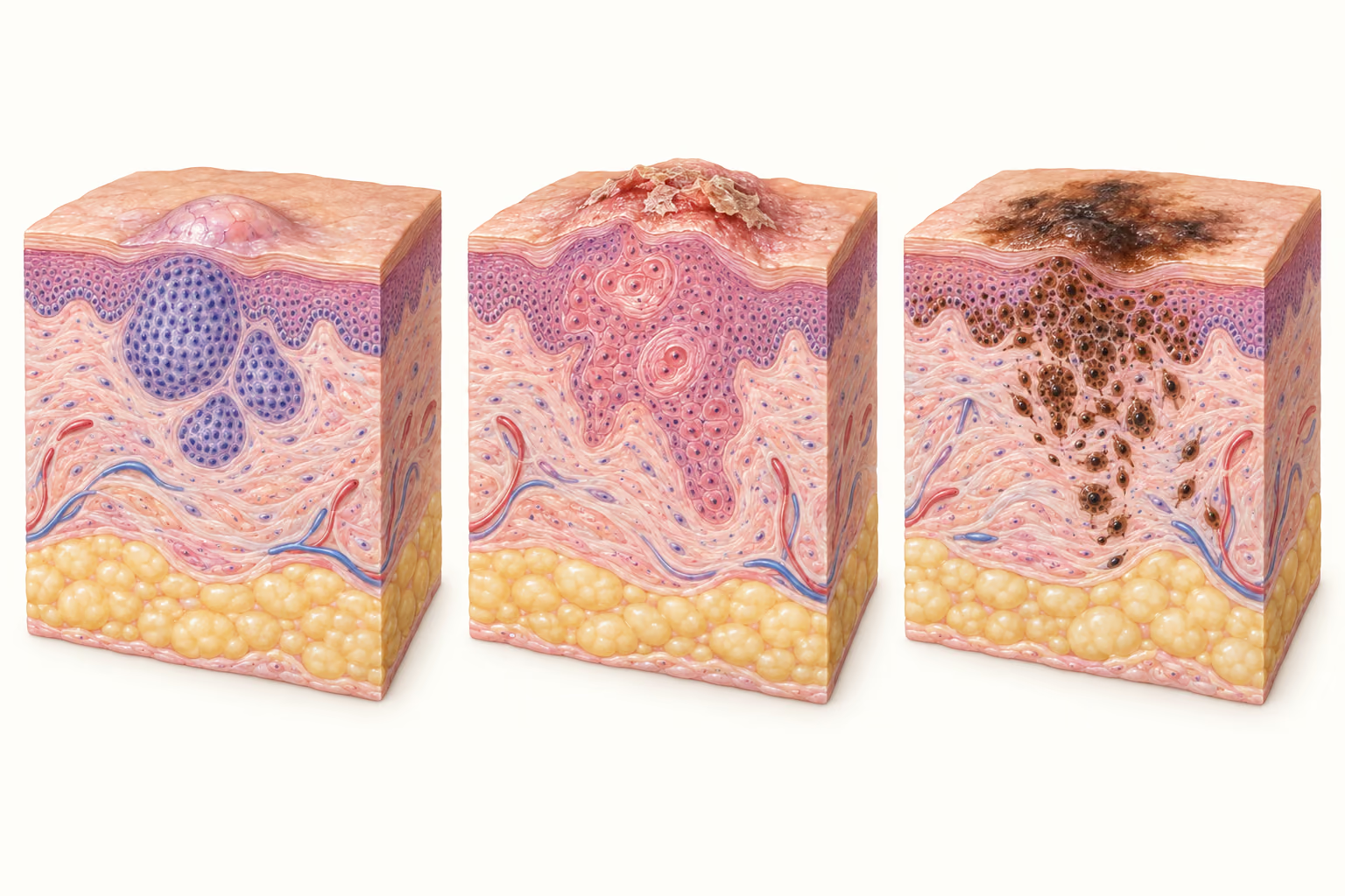
Educational comparison: appearance alone does not establish the diagnosis.
Type
Behaviour
Treatment considerations
Basal cell carcinoma (BCC)
Often grows slowly and locally. It may ulcerate and invade nearby tissue; metastasis is exceptionally rare.
Site, size, subtype, borders and recurrence. Complete removal is the main option for many cases.
Squamous cell carcinoma (SCC)
May be confined to the epidermis (in situ) or invasive. Some tumours have a higher risk of involving nerves, lymph nodes or other organs.
Depth, differentiation, perineural invasion, location, recurrence and immunosuppression.
Melanoma
Arises in melanocytes and can spread. Thickness and other pathology findings are particularly important.
Histological diagnosis, Breslow thickness, ulceration, stage and possible lymph-node assessment.
A red or lightly pigmented lesion can also be malignant. Skin cancer occurs in different skin tones and may affect areas with little sun exposure. Internet photographs cannot replace an examination and biopsy.[1][2][4]
AI-generated conceptual illustration: left to right, basal cell carcinoma, squamous cell carcinoma and melanoma. Not to scale and not a self-diagnosis tool.
03 / SKIN CANCER GUIDE
What can happen if treatment is delayed?
A small lesion may require a larger operation later.
As a tumour grows, removal may involve more tissue and more complex reconstruction. Bleeding, ulceration and loss of function may also occur. Risk varies: diagnosis and risk assessment determine priority.[1]
BCC: damage is usually local
The rarity of metastasis does not make BCC harmless. Persistent tumours can damage important structures, especially around the eyes, nose and ears. Recurrence and aggressive subtypes influence the surgical approach.[5]
SCC: risk assessment matters
Invasive SCC can progress beyond the skin. Immunosuppression, nerve involvement and certain pathological findings increase concern and may require specialist investigation and treatment.[6][7]
Melanoma: stage changes management
A melanoma that becomes thicker or reaches lymph nodes or other organs may need treatment beyond local removal. Investigating a suspicious lesion and completing the recommended treatment avoids losing important time.[2]
Missing the pathology appointment
A healed wound does not prove treatment is complete. Involved margins or high-risk findings may require further surgery, tests or referral. Agree how you will receive and discuss the report.
A photograph cannot establish how many days a lesion can safely wait. If access to recommended assessment or surgery is difficult, tell the care team so they can help plan a route to treatment.
04 / SKIN CANCER GUIDE
Before removal: examination, dermoscopy and biopsy
The consultation reviews how long the lesion has been present, changes over time, sun exposure, previous treatments, other skin cancers and medication. Dermoscopy helps assess skin structures, but does not replace histology when a biopsy is indicated.
A biopsy removes part or all of a lesion for microscopic examination. The technique depends on the suspected diagnosis, size and location. The sample needs to provide enough information for the next decision; an unsuitable superficial sample can make depth assessment difficult.[8][2]
When melanoma is suspected, planning aims to preserve assessment of the whole lesion and its thickness. Sampling is tailored for certain anatomical locations or extensive lesions. One technique is not appropriate for every skin mark.[9]
A suspicious lesion should not be destroyed before its diagnosis is clarified.
Acids, cautery and other destructive methods may prevent adequate tissue examination. Electrocautery has different indications from planned cancer excision with margin assessment. Do not attempt home removal.
Surgery aims to remove the tumour with cancer control, preserve function and achieve the best possible repair. Technique, anaesthesia and setting depend on the individual case.
01
Planning
The diagnosis and site are reviewed along with risk, medication and alternatives. The visible size of a lesion does not by itself determine how much tissue needs removal.
02
Anaesthesia and removal
Many skin operations use local anaesthesia. Extensive disease or particular patient needs may require additional support and a hospital setting.
03
Tissue examination
The labelled specimen is sent for pathology. The chosen technique determines how and when the margins will be examined.
04
Repair and review
Direct closure, a flap, a graft or healing by secondary intention may be appropriate. Follow-up covers the wound, pathology and further care.
Conventional excision
The tumour is removed with a surrounding border of tissue determined by clinical criteria. Routine microscopic assessment occurs after surgery. Involved margins may require additional treatment.[1]
Mohs micrographic surgery
Tissue is removed in stages, with mapping and examination of margins during the procedure. It may be appropriate for selected high-risk or recurrent tumours and sites where preserving tissue is particularly important.[1][5]
Mohs requires a dedicated team and facilities. Indications and referral are discussed at assessment; describing the technique here does not imply that it is available at this clinic.
06 / SKIN CANCER GUIDE
Surgical margins and pathology: why do they matter?
The clinical margin is the border planned by the surgeon around a tumour. The histological margin is the edge of the removed tissue examined by the pathologist. These concepts are related, but they are not the same measurement. An internet rule cannot determine the right margin for an individual patient.
Clear margins mean that no tumour was found at the examined edges. This is an important finding, but it does not eliminate every risk of recurrence or replace follow-up. Interpretation also depends on the specimen and examination method.
Involved or positive margins mean tumour reaches an examined edge. Further excision, margin-controlled surgery or another approach may be recommended according to the tumour and clinical context. A normal-looking scar does not exclude residual tumour.[1][10]
Type and subtype
Identify the tumour and features that matter for risk assessment.
Depth and invasion
Describe extension into tissue and possible involvement of structures such as nerves.
Breslow thickness and ulceration
In melanoma, these help determine stage and guide surgery and additional testing.
A documented plan
The report should be discussed with the care team and followed by a clear management plan.
Request a copy of the report. If the laboratory's expected turnaround has passed, contact the team: no message should not be interpreted as a normal result.[2][8]
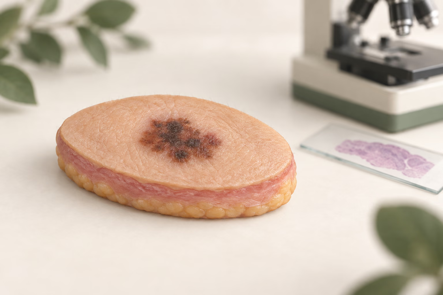
AI-generated educational model of excised tissue with a lesion and surrounding border. Only examination of the specimen can assess the margins; appearance does not prove clearance.
07 / SKIN CANCER GUIDE
Melanoma: removing the mole may only be the first step
After a biopsy confirms melanoma, treatment may include wider excision around the scar even when the visible lesion has already been removed. The purpose and extent of further surgery depend on the pathology and stage.
Breslow thickness is a microscopic measurement of invasive melanoma depth. It is not the mole's diameter. Ulceration and other findings add to the assessment. “In situ” means melanoma remains confined to the epidermis; it still requires appropriate treatment.[2]
Sentinel lymph node biopsy is a staging test considered in selected situations after discussing its benefits and risks. It is not required for every melanoma and does not automatically mean removing all lymph nodes. Its indication should be coordinated with definitive surgery.[9][2]
Higher-risk, regional or advanced disease may require oncology care, imaging and systemic treatment such as immunotherapy or targeted therapy in specific settings. In some situations medication is considered before surgery. These cases need multidisciplinary planning.[2]
08 / SKIN CANCER GUIDE
Is surgery always the only option?
Surgery is central to treating many localised skin cancers. However, decisions consider diagnosis, recurrence risk, general health, site and informed patient preferences.
Topical treatment, photodynamic therapy and destructive methods have a role in certain superficial or low-risk lesions. Radiotherapy may be considered in selected circumstances. These treatments are not interchangeable with surgery and should not be used to obscure a suspected melanoma.[1][5]
Systemic options may be available for advanced disease or when surgery is unsuitable. Availability, indications and risks need discussion with the treating team. The decision should compare tumour control, adverse effects, possible additional procedures and follow-up.[1][2]
09 / SKIN CANCER GUIDE
Preparing for assessment and surgery
Bring to the consultation
Pathology reports, tests, earlier photographs and details of previous surgery at the same site.
A medication and supplement list, allergies and previous anaesthetic reactions.
Information about diabetes, immunosuppression, transplantation, bleeding and past wound healing.
Questions about work, transport, dressings, costs and follow-up.
Agree before surgery
Which procedure is planned and why it was selected.
The setting, anaesthesia, any fasting requirements and whether a companion is needed.
How the wound will be repaired and which activities will be restricted.
Who will communicate pathology results and when you should return.
Do not stop anticoagulants or other medicines on your own.
Bleeding risk must be weighed against the risk of stopping treatment. Any adjustment should be agreed with the surgeon and, when needed, the prescribing clinician. Also report a pacemaker or other implanted device.[11]
Costs are individual: assessment, excision size, reconstruction, laboratory analysis and surgical setting may affect the plan. Ask what is included and whether another team will be involved.
10 / SKIN CANCER GUIDE
Surgical risks to discuss
The expected benefit of removal must be weighed against procedural risks and the risks of leaving the cancer untreated. No surgery is complication-free, and consent should address the particular lesion and your health.
Bleeding and infection
These may require assessment, extra dressings or treatment. Report increasing pain, discharge or spreading redness.
Wound separation and slow healing
Tension, location, circulation and medical conditions influence recovery.
Scarring and altered sensation
Marks, colour changes, numbness or injury to nearby structures can occur, depending on the site.
Reconstruction and further procedures
Flaps and grafts have their own risks. Pathology findings or recurrence may also lead to another operation.
The frequency of complications depends on the operation and patient; one percentage for every skin operation would be misleading.[11][1]
11 / SKIN CANCER GUIDE
Recovery: wound care and completing treatment
Before leaving
Obtain written instructions for dressings, washing, medication and contacting the team about problems. Confirm activity restrictions and follow-up.
During healing
Follow the prescribed cleansing and dressing plan. Do not use home remedies or remove stitches yourself. Flaps and grafts may need different care from a small biopsy wound.
At follow-up
Review the wound and pathology. Stitch removal and return to exercise, swimming or the sea depend on the site and healing; there is no universal schedule.
If symptoms worsen
Persistent bleeding, fever, discharge or increasing pain need contact with the team. Seek urgent care for heavy bleeding or feeling seriously unwell.[3]
Tumour removal, wound healing and cancer control are connected but distinct parts of care. One does not replace the others.
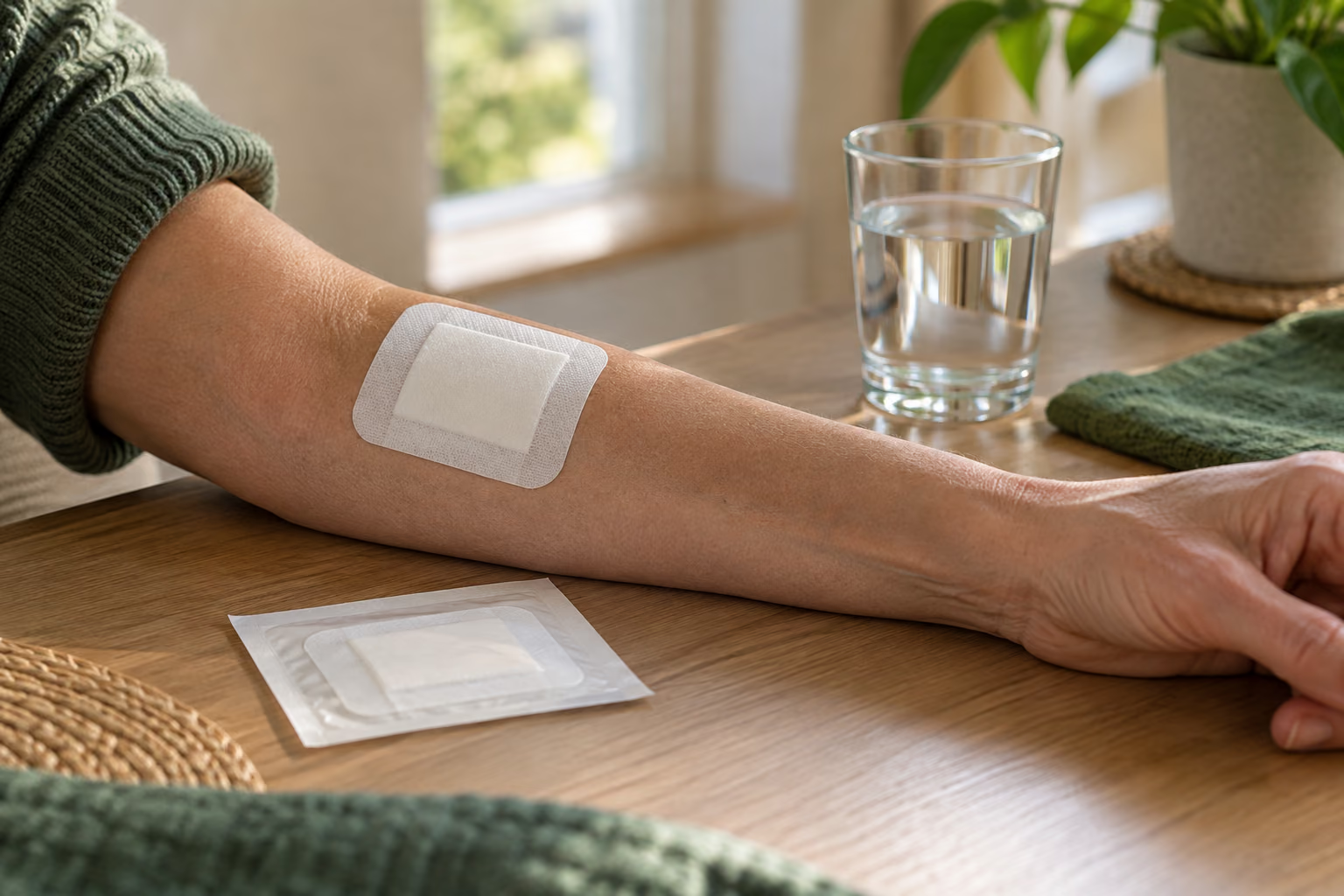
AI-generated illustrative scene of an arm with a dressing. Wound care and dressing selection should follow individual clinical instructions.
12 / SKIN CANCER GUIDE
Follow-up after removal
Follow-up looks for recurrence, new tumours and scar-related needs. Appointment frequency depends on diagnosis and risk; melanoma, multiple tumours or immunosuppression may require a more specific plan.
Check your skin between visits and report new lesions, scar changes or persistent lumps near the treated area. Do not wait for a scheduled appointment if a concerning change appears. Organised photographs can help show evolution but do not replace examination.[1][2]
Sun protection remains important: seek shade, use suitable clothing and a hat, and follow advice on sunscreen. Outdoor work deserves a practical protection plan. Treating one tumour does not remove the possibility of developing another.[4]
13 / SKIN CANCER GUIDE
What published research adds to the decision
European guideline · 2023
BCC treatment according to risk
The European consensus prioritises complete surgery and recommends micrographic control in higher-risk settings. Nonsurgical alternatives are reserved for selected situations. Guidance addresses patient groups and does not replace individual assessment.[5]
Randomised trial · 10 years
Recurrence may occur late
In a trial of high-risk facial BCC, recurrent tumours had ten-year recurrence rates of 3.9% after Mohs and 13.5% after conventional excision. For primary tumours, the difference was not statistically significant. These figures are not a guaranteed success rate for every lesion and support long-term follow-up.[12]
Clinical guideline · SCC
Pathology helps identify high risk
AAD recommendations combine clinical and histological features to guide treatment and surveillance. A pathology result should not be reduced to the word “cancer” alone.[6]
ESMO guideline · 2025
Melanoma needs stage-based planning
The guideline addresses diagnosis, treatment and follow-up, including stage-based decisions on surgery and systemic therapy. Application in Brazil also depends on clinical assessment and access to indicated treatments.[9]
Selected guidelines, institutional information and clinical research consulted on 17 September 2026. Full references and article DOIs appear below. Educational content: individual care is determined at consultation.
QUESTIONS FOR YOUR CONSULTATION
40 questions about skin cancer surgery
No questions found. Try another word.
01Is every bleeding lesion skin cancer?
No. Trauma and benign conditions can also bleed. However, spontaneous or repeated bleeding from a persistent lesion warrants assessment. Appearance alone cannot confirm or exclude cancer.[1]
02Should a sore that does not heal be assessed?
Yes. A persistent sore, or one that seems to heal and then reopens, needs examination. Avoid repeatedly treating it with creams without identifying the cause, especially if it grows.[1]
03Does skin cancer always hurt?
No. A malignant lesion may be painless. Lack of pain does not establish that a mole or sore is benign; changes and examination guide investigation.[1][2]
04Can a small mole be melanoma?
Yes. Size is only one feature assessed. Change, asymmetry, different colours or a mole unlike the others warrants assessment even when it is small.[2]
05Can people with dark skin develop skin cancer?
Yes. People of every skin tone can develop skin cancer. Do not ignore changes on palms, soles, nails or areas with little sun exposure.[2]
06Is a dark nail stripe always melanoma?
No. There are benign causes. A new or changing stripe, especially with nail changes or pigment around it, should be examined; a description alone cannot establish the diagnosis.[2]
07Can BCC be left untreated because it grows slowly?
Slow growth does not mean absence of risk. The tumour may cause local destruction. Treatment, or any exceptional decision to observe, needs individual assessment and a documented plan.[1]
08Can squamous cell carcinoma spread?
Yes, although risk varies. Pathology and examination help identify high-risk tumours and determine whether lymph nodes or other areas need investigation.[1]
09Does removal cure every case?
There is no universal guarantee. Many localised tumours can be controlled with appropriate treatment, but outcomes depend on type, stage, margins and other factors. Some cases need additional treatment.[1][2]
10Must a biopsy precede every operation?
Histology is part of diagnosis, but the sequence varies. Initial complete removal may also serve as the biopsy; sometimes an earlier sample is needed to plan treatment.[1][2]
11Does dermoscopy replace biopsy?
Not when tissue analysis is indicated. Dermoscopy adds information to examination; pathology identifies microscopic features needed for diagnosis and planning.[1][2]
12Are biopsy and definitive removal the same?
Not always. A biopsy establishes the diagnosis. Even after removal of all visible disease, the result may indicate wider excision or another treatment. Ask about the procedure's purpose.[1][2]
13Can a suspicious mole be cauterised?
A suspicious lesion needs diagnosis before a destructive method is chosen. Cautery may compromise tissue analysis and does not replace planning for cancer surgery.[1][2]
14How is the surgical margin chosen?
It depends on type, size, location, subtype and risk. In melanoma, pathology also guides wider excision. Another patient's margin is not a prescription for your case.[1][2]
15What does a clear margin mean?
No tumour was found at the examined edges. This is favourable, but it must be interpreted with the rest of the report and does not remove the need for follow-up.[1]
16Does an involved margin require another operation?
It often prompts discussion of further treatment. The decision considers type, site, risk and health. A healed wound does not prove the absence of residual disease.[1]
17Is Mohs suitable for every skin cancer?
No. It is important in selected situations. Type, risk, recurrence and location help determine its benefit. Referral to a service with dedicated facilities may be needed.[1]
18Does the clinic perform Mohs surgery?
Availability must be confirmed at assessment. This guide explains options and does not advertise Mohs at this clinic. The plan may include specialist referral when appropriate.
19Is skin cancer surgery painful?
Anaesthesia aims to control pain. There may be discomfort during administration and afterwards. Tell the team if you feel pain and use only recommended medication.[8]
20Will I need hospital admission?
Many procedures are outpatient operations, but not all. Tumour extent, reconstruction, anaesthesia and health determine the appropriate setting.[1]
21Can the scar be larger than the tumour?
Yes. Removal includes surrounding tissue, and closure may require a larger outline than the visible lesion. The aim is to balance tumour control, function and repair.[1]
22When is a flap or graft needed?
These may be needed when simply bringing the wound edges together is unsuitable. Site, size, depth and tissue condition guide reconstruction.[1]
23Should I stop my anticoagulant before surgery?
Not on your own. The team must assess bleeding risk and the risk of stopping medication. Report all medicines and follow the agreed plan.[11]
24Do diabetes or immunosuppression rule out surgery?
Not necessarily. They may change risk and care requirements. Report diabetes control, transplantation and immunosuppressive medicines so planning reflects your needs.[11][1]
25How long does pathology take?
Turnaround varies by laboratory and whether additional analysis is required. Confirm the expected time and who will communicate the result. Contact the team if it is delayed.
26The wound has healed: can I skip the pathology review?
No. Appearance does not reveal tumour type, margins or other microscopic findings. The report needs review even if the wound looks fully healed.[1][2]
27When can I return to work and exercise?
It depends on the surgical site, repair and demands of the activity. Agree individual restrictions for exertion, lifting, friction and movements that put tension on the wound.
28Can a scar change during recovery?
Its appearance can evolve during healing. However, a new lump, persistent sore or progressive change at the treated site needs assessment; do not automatically attribute every change to scarring.[1]
29Can skin cancer return at the same site?
Yes. Recurrence differs from a new tumour elsewhere. Risk depends on the disease and treatment, so follow-up remains important.[1][2]
30Can someone who has had skin cancer develop another?
Yes. Treating one lesion does not remove the risk of new tumours. The plan may include regular skin examinations, sun protection advice and self-observation.[1]
31Does every melanoma require lymph-node removal?
No. Sentinel-node assessment is indicated in selected situations. Even a positive result does not automatically mean removing all lymph nodes; management depends on stage and specialist assessment.[2]
32What is Breslow thickness?
It is a microscopic measurement of invasive melanoma depth, not the width of the mole. Alongside other findings, it helps guide staging and treatment.[2]
33Does melanoma in situ need treatment?
Yes. Being confined to the epidermis does not mean it is benign. The aim is appropriate treatment, usually with surgery planned for this diagnosis.[2]
34Why have wider surgery if the mole is already gone?
The initial removal may have been diagnostic. Pathology may indicate a different definitive margin. For melanoma, wider excision is planned according to histological findings.[2]
35Will I need chemotherapy or immunotherapy?
Not every skin cancer needs systemic treatment. Type and stage guide this decision. For higher-risk or advanced disease, the team may discuss immunotherapy, targeted therapy or other options.[1][2]
36Can a cream replace surgery?
In specific settings, topical treatments are options for certain superficial, low-risk lesions. They are not suitable for every tumour and should not be used without diagnosis and follow-up.[1]
37Does every case require a CT scan?
No. Imaging depends on tumour type, stage, symptoms and clinical findings. Deciding which tests are needed is part of individual planning.[1][2]
38Can I wait a few months for treatment?
This guide cannot establish a safe waiting time. If cancer is suspected or confirmed, seek assessment and follow the priority set by the care team. Report difficulty accessing treatment.[1][2]
39How much does skin cancer removal cost?
Cost depends on assessment, technique, size, reconstruction, laboratory analysis and surgical setting. Request an individual estimate and clarification of what is included; visible lesion size alone is insufficient.
40How can I prepare for an assessment in São Mateus, ES?
Contact the clinic and mention any established diagnosis or biopsy. Bring reports, medication details and records of changes. Assessment determines indications, technique, treatment setting and any referral.
NEXT STEP
Skin lesion assessment in São Mateus, ES
The first step is to clarify the diagnosis and choose appropriate treatment. Bring existing tests and pathology reports. Surgical indications, the required setting and any referral depend on assessment.
Prepare for your consultation with Dra. Caroline Minchio
When contacting the clinic, mention changes in the lesion and any established skin cancer diagnosis. Digital channels help with booking; they do not replace emergency care.
References follow ABNT NBR 6023:2025 in order of first citation. Original titles are retained, with publication details, DOI and access date where applicable.
PERIS, K. et al.. European consensus-based interdisciplinary guideline for diagnosis and treatment of basal cell carcinoma—update 2023. European Journal of Cancer, [S. l.], v. 192, art. 113254, out. 2023. DOI: 10.1016/j.ejca.2023.113254. Disponível em: https://pubmed.ncbi.nlm.nih.gov/37604067/. Acesso em: 17 set. 2026.
KIM, J. Y. S. et al.. Guidelines of care for the management of cutaneous squamous cell carcinoma. Journal of the American Academy of Dermatology, [S. l.], v. 78, n. 3, p. 560–578, mar. 2018. DOI: 10.1016/j.jaad.2017.10.007. Disponível em: https://pubmed.ncbi.nlm.nih.gov/29331386/. Acesso em: 17 set. 2026.
AMARAL, T. et al.. Cutaneous melanoma: ESMO Clinical Practice Guideline for diagnosis, treatment and follow-up. Annals of Oncology, [S. l.], v. 36, n. 1, p. 10–30, jan. 2025. DOI: 10.1016/j.annonc.2024.11.006. Disponível em: https://pubmed.ncbi.nlm.nih.gov/39550033/. Acesso em: 17 set. 2026.
VAN LOO, E. et al.. Surgical excision versus Mohs’ micrographic surgery for basal cell carcinoma of the face: a randomised clinical trial with 10 year follow-up. European Journal of Cancer, [S. l.], v. 50, n. 17, p. 3011–3020, nov. 2014. DOI: 10.1016/j.ejca.2014.08.018. Disponível em: https://pubmed.ncbi.nlm.nih.gov/25262378/. Acesso em: 17 set. 2026.
Educational content updated on 17 September 2026. It does not replace consultation, histological diagnosis or an individual treatment plan. No cure or cosmetic outcome is guaranteed.