Bleeding or infection
Bleeding that persists despite pressure, worsening severe pain, pus with spreading redness or fever needs assessment. Heavy bleeding or marked illness warrants urgent care.[3] [4]
SURGICAL DERMATOLOGY · SÃO MATEUS, BRAZIL
Has a lesion changed or started bleeding? Arrange a diagnostic assessment. Cautery is considered only after the cause is understood. See when to seek care.
Small skin lesions can cause daily discomfort. Understand when treatment may help, what waiting can mean and how to choose care suited to your skin.
Seek assessment. These signs may require investigation before any cautery treatment.[1] [2] See when to seek care ↓
Dra. Caroline Minchio · CRM-ES 15578 · São Mateus, Espírito Santo, Brazil.
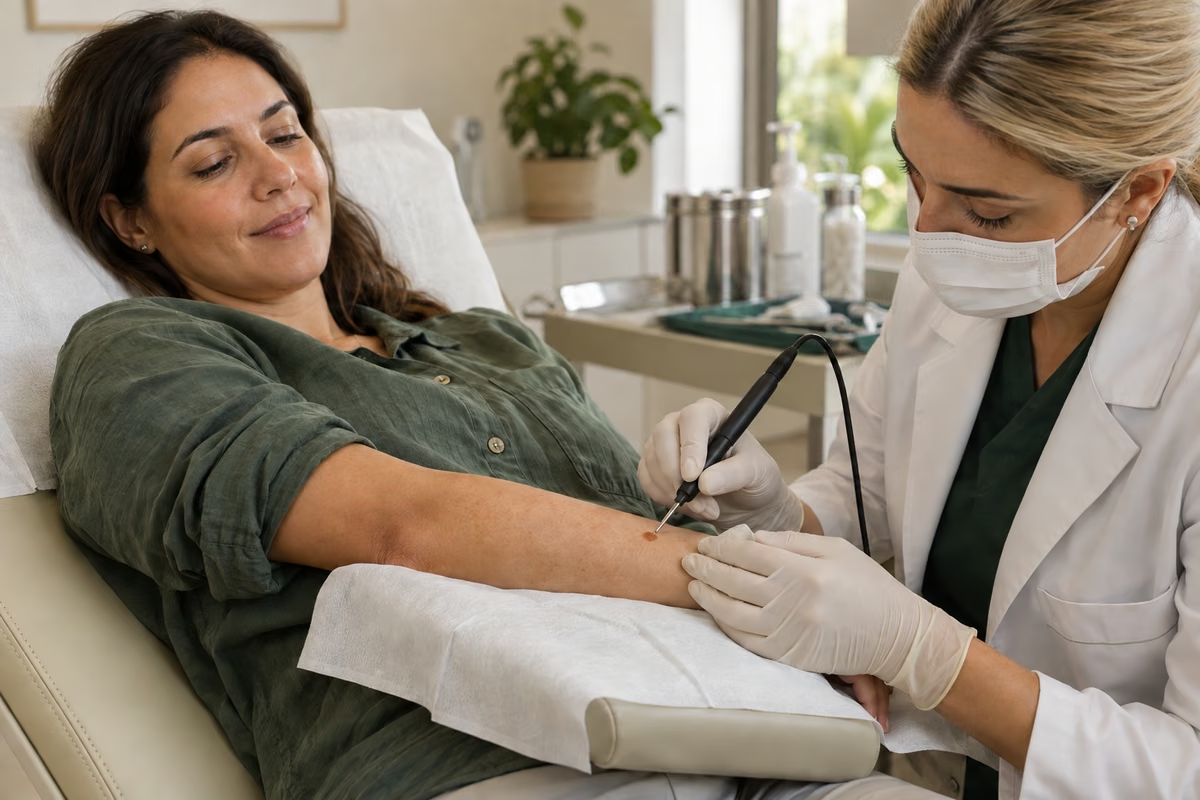
Assess · choose · treat · follow up
THE RIGHT TIME FOR CARE
Signs and diagnosis determine priority. Needing prompt assessment does not mean cautery on the same day will be the right treatment.
Bleeding that persists despite pressure, worsening severe pain, pus with spreading redness or fever needs assessment. Heavy bleeding or marked illness warrants urgent care.[3] [4]
Rapid growth, changing shape or colour, spontaneous bleeding or a non-healing sore deserves investigation. Do not wait for a convenient cosmetic treatment date.[1] [2]
An assessed, stable lesion without warning signs may allow planned treatment or monitoring. Irritation, discomfort and personal preferences inform the decision.[5] [6]
WHAT WAITING CAN MEAN
Lesions caught by clothing, jewellery or a razor may remain irritated, painful or prone to bleeding. Repeated problems are a reason to discuss planned treatment.[5] [6]
Viral warts can persist and appear elsewhere. Treatment may help, but the technique should suit the site and the individual.[7]
Ignoring a suspicious lesion can delay investigation. Burning a lesion before clarifying the diagnosis may also destroy tissue needed for analysis.[1] [2]
UNDERSTANDING THE TECHNIQUE
It is a technique that uses heat to treat tissue or control small areas of bleeding. In everyday language, the term is also used for electrosurgery. Technically, electrocautery heats a tip, while electrosurgery uses high-frequency electrical energy to produce a tissue effect.[8]
The doctor decides the method, extent and depth after examining the skin. Laser, cryotherapy and other devices work differently. Suitable selection and healing care are central to the benefit.
WHEN IT MAY HELP
A similar appearance does not guarantee the same diagnosis. Examples in which this technique may be considered:
Small skin growths that may become irritated by friction. Removal may help with discomfort or appearance after diagnosis is confirmed.[5]
Benign growths with varied surfaces. Electrosurgery, curettage, cryotherapy and other options are weighed against thickness, location and pigment-change risk.[6] [9]
Several treatments are available. Location, number, recurrence, immunity and sensitivity of the area influence whether electrosurgery is appropriate.[7]
Selected small vascular lesions or sebaceous hyperplasia may be considered. The technique does not replace assessment of a bleeding or changing spot.[10]
FROM ASSESSMENT TO FOLLOW-UP
Examine the lesion, discuss its history and decide whether dermoscopy or tissue sampling is needed.
Compare techniques, explain risks, discuss expectations and obtain consent.
Prepare the area, provide appropriate anaesthesia and perform the procedure in a suitable setting.
Receive care instructions, learn how to obtain any pathology report and arrange review.
This describes the care pathway, not instructions for performing the procedure.[13] [1]
What is the lesion? Why treat now? Which technique fits? What care and timing will be needed? Bring these questions to your assessment.
A SHARED DECISION
No technique is best for every lesion. This comparison helps organise the discussion.
| Option | When to discuss it | Key consideration |
|---|---|---|
| Monitoring | Confirmed benign lesion without significant symptoms. | Agree which changes require reassessment. |
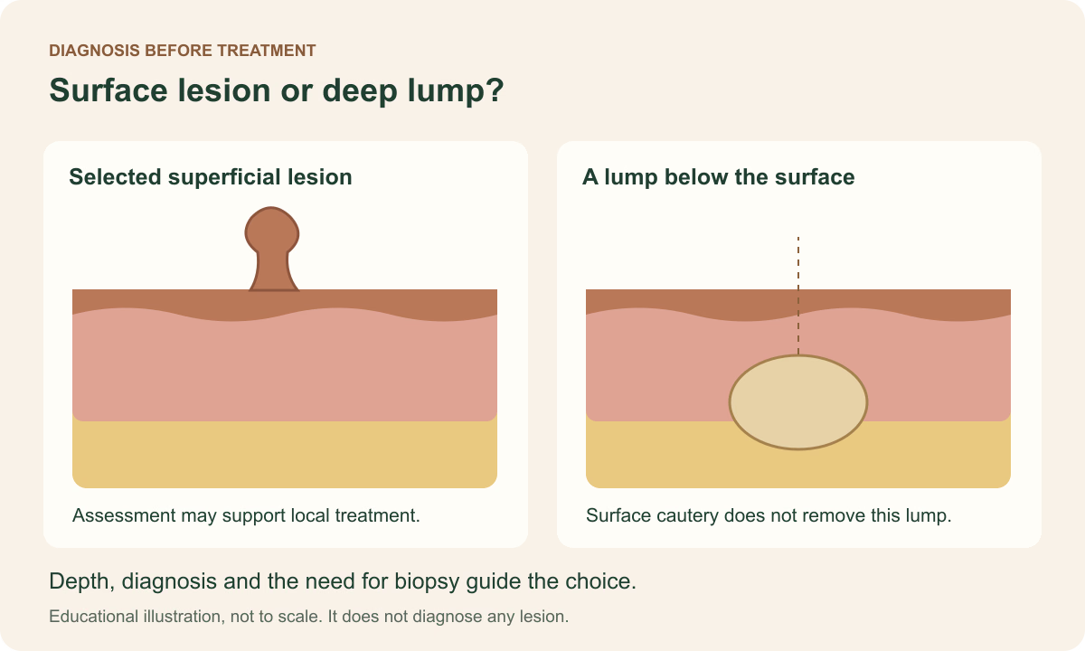
| Electrosurgery / cautery | Selected superficial lesions and bleeding control. | Scars or pigment changes are possible; plan sampling before destroying tissue. |
| Cryotherapy | Warts and other selected lesions. | May require repeat treatment and cause blisters or pigment changes. |
| Biopsy / excision | Diagnostic uncertainty or a need for removal and analysis. | Technique and closure depend on depth and location. |
Other options, including topical treatments or laser, may be appropriate depending on diagnosis.[5] [7] [6] [1] [12]
BENEFITS AND LIMITATIONS
Even a small procedure creates a wound and needs care. Individual assessment helps reduce risks and choose appropriate timing.
Pale or dark marks, depressions or raised scars may occur. Previous keloids and skin type should be considered.
The site and medicines affect risk. Increasing pain, pus or progressive redness after treatment needs assessment.
Heat can extend beyond the intended area. Equipment, technique, protection and communication all contribute to safety.
Reassessment, repeat treatment or a different approach may be needed. A returning lesion should not be repeatedly cauterised without reviewing the diagnosis.
RECOVERY IS PART OF TREATMENT
Confirm dressing care, cleansing, permitted products, follow-up and a contact for questions. Ask for written instructions.
Follow your care plan and protect against friction. Do not pick scabs, scrub or add acids or home remedies to the wound.
Clearance for exercise, swimming and makeup depends on the site and healing. Colour changes can last longer than wound closure.
Protect from sun and compare healing with the team’s expectations. If tissue was analysed, obtain the result and agree on the next step.
Your procedure-specific instructions take precedence over this general guide.[13] [3]
HOW RESEARCH INFORMS THE DECISION
A study of 33 participants compared cryosurgery and electrodesiccation in similar lesions. Its design helps compare options in that setting; it does not establish one best technique for all lesions.
This study compared cryotherapy and electrosurgery in four types of benign lesions. Results differed by lesion type, and both groups experienced adverse effects. Individual selection remains important.
Studies are not promises of results. Sample, technique, skin type and follow-up limit their application to an individual. Institutional guidance complements research with diagnostic and care information.
ANSWERS TO SUPPORT YOUR DECISION
No matching question. Try another word.
It uses heat to treat tissue or control bleeding. Ask which technique is planned: electrocautery and electrosurgery are not technically identical.[8]
No. These are different technologies. Diagnosis, depth, location and realistic outcomes guide the choice; the device name alone does not determine the best treatment.[8] [6]
Selected skin tags, seborrhoeic keratoses, warts and other superficial lesions. Examination comes before treatment; spots, moles and lumps are not interchangeable diagnoses.[13] [5] [7]
Urgency depends on the lesion. Suspicious changes need prompt assessment; stable benign lesions may allow planned treatment. Persistent bleeding or infection signs need timely care.[2] [4]
Irritation or bleeding may continue; warts may spread. With a suspicious lesion, delayed diagnosis is the key concern. Confirmed benign lesions can sometimes be monitored.[5] [7] [2]
No. Diagnosis, symptoms, location, preferences and treatment risks all matter. A harmless lesion that causes no trouble may not need a procedure.[5] [6]
Yes. Different lesions can look alike. Rapid growth, ulceration, spontaneous bleeding or colour change needs examination, rather than an attempt at home cautery.[7] [2]
A mole needs its own assessment. Destroying suspicious tissue can compromise analysis. The doctor may choose a biopsy or removal that preserves a specimen.[1] [2]
It depends on the lesion and technique. If analysis is needed, sampling must be planned before tissue is destroyed. Ask how and when you will receive the report.[13] [1]
Curettage with cautery can suit selected low-risk tumours. It is not a universal treatment and does not replace diagnosis, staging or choosing the appropriate technique.[11]
Local anaesthesia is commonly used and its injection may sting. Tell the clinician if you feel pain. Afterwards, some tenderness is possible.[13]
Many small procedures use local anaesthesia. The extent, location, age and health of the patient guide planning; cases differ.[13]
The intended effect is localised to the tissue. The technique and anaesthesia are chosen for the procedure. Tell the team immediately about any painful sensation.[8] [13]
Duration depends on the number, size and location of lesions, anaesthesia and any sampling needed. Ask for an estimate during assessment.
Sometimes, after assessment and consent. Biopsy planning, preparation, medicine review or scheduling may require another date.
There is no single number. Some lesions need one session; others need review, additional treatment or a different technique. Agree on follow-up.[7] [6]
Persistence or recurrence is possible, especially with warts. New lesions can also develop elsewhere. A returning lesion should be reassessed.[7]
A pale, dark, flat or raised mark is possible. Depth, location, individual healing and aftercare matter. Scar-free skin cannot be guaranteed.[13] [4]
People who are prone to them can develop raised scars. Report previous keloids or thick scars; this affects the discussion of risks and alternatives.[13] [4]
Pigment changes deserve particular attention. Skin type, previous discolouration and location should be considered; another technique may sometimes be preferable.[6] [10]
Tell the team about a pacemaker, defibrillator or other implanted device before scheduling. Device identification and technique planning may require cardiology input.[8] [14]
Do not stop them on your own. Bring a complete medicine list; bleeding risk must be weighed against the risk of interrupting treatment.[4]
It can affect healing and infection risk. Discuss glucose control, medicines and past wound problems, especially on the feet.[7] [4]
Tell the team. The need, timing, anaesthesia and medicines require individual assessment. Cosmetic treatment may be planned for another time.
These sites require specific assessment and protection. Diagnosis and technique may differ; this guide is not a substitute for advice on genital or near-eye lesions.[5] [7]
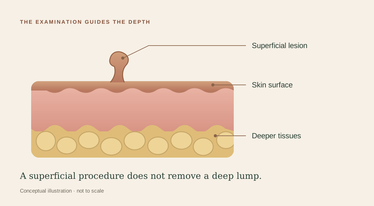
Cauterising the surface is not equivalent to removing a subcutaneous mass or a cyst capsule. These conditions need their own assessment and treatment.
Curettage and cautery commonly heal without stitches. If excision is also needed, closure may differ. Confirm which procedure is planned.[13]
Timing varies with location, extent and depth. Some superficial wounds close within a few weeks; the appearance of the mark continues to change afterwards.[13] [3]
Follow the timing and washing instructions from your team. Gentle cleaning is often part of care; do not scrub or add irritating products on your own.[3]
Do not pick or pull at it. Protect the area as advised and report worsening rather than trying to force healing.[3]
Use only what your clinician recommends. A topical antibiotic is not required for every wound and can cause allergy. Prescribing depends on the case.[3]
Avoid applying them to an open wound. Ask the team when to restart makeup, retinoids, acids and other products on that area.
Protect the area from sun and ask when immersion is safe. Clearance depends on healing. Follow advice about sunscreen once the skin has closed.[3]
It depends on the treated site, friction, sweating and exertion involved. Ask for instructions tailored to your routine before leaving.
Increasing pain, spreading redness, pus, fever or persistent bleeding requires contact with the team and assessment. Marked illness needs urgent care.[3] [4]
Thermal injury is a risk. Appropriate technique, equipment and protection reduce but do not eliminate it; depth must be controlled by the clinician.[8]
No. In addition to burns, scarring and infection, destroying an undiagnosed lesion can delay recognition of an important condition.[8] [1] [4]
The estimate depends on assessment, lesion number and size, the technique and any pathology. Ask what is included and how follow-up works.
No. Studies involve specific groups, techniques and follow-up periods. Evidence informs choices but cannot predict your individual scar, recurrence or recovery.[9] [10]
What is the diagnosis? Why this technique? Do I need a biopsy? What if I wait? What marks may remain? When is follow-up, and whom do I contact if there is a problem?
BIBLIOGRAPHY AND SOURCES
Sources consulted on 17 September 2026. References follow ABNT NBR 6023:2025 and are ordered by first citation. They include clinical studies, a professional guidance update and institutional guidance.
Educational information. Diagnosis, treatment selection and timing depend on individual assessment. Images cannot be used for self-diagnosis.
KEEP READING
Understand biopsy, removal and the risks of delaying treatment →
Understand the differences, indications, risks and aftercare of these procedures.