Growth and discomfort
Some lipomas enlarge and cause pain, pressure or interference with daily activities. Delaying removal recommended for these symptoms can prolong discomfort. Large or deep lesions may need more complex surgical planning.[7] [11]
DERMATOLOGY GUIDE · SÃO MATEUS, BRAZIL
A confirmed lipoma can often be monitored. Understand the risks of ignoring a changing lump, when removal may help and what to consider before surgery.
Rapid growth, persistent pain or a hard, fixed lump warrants prompt assessment.
Understand the risks and treatment options ↓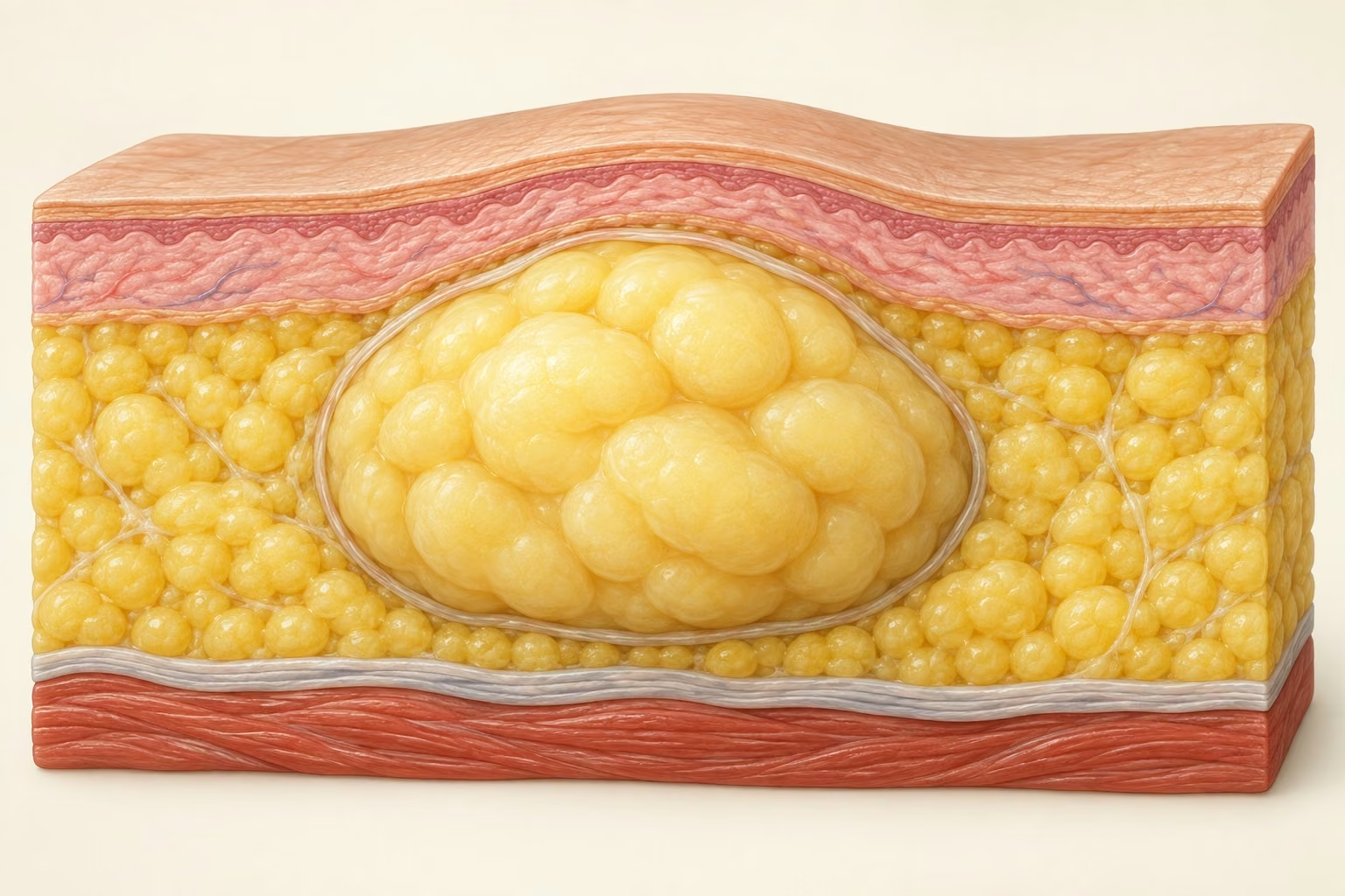
INFORMATION FOR YOUR DECISION
The decision begins with a diagnosis. A lump labelled a “lipoma” at home still needs examination. Monitoring or surgery then depends on its symptoms and clinical features.
Some lipomas enlarge and cause pain, pressure or interference with daily activities. Delaying removal recommended for these symptoms can prolong discomfort. Large or deep lesions may need more complex surgical planning.[7] [11]
Ignoring a rapidly growing or unusual mass can delay investigation of another cause, including a tumour needing specialist care. An ordinary lipoma is benign; the aim is to confirm whether the mass really is a lipoma.[3] [4]
Removal may be considered for growth, pain, functional problems or significant discomfort after diagnostic assessment. A suspicious mass needs planned investigation and specialist referral before biopsy or removal.[3] [7] [11]
Small, stable lipomas without symptoms can often be monitored after assessment. Agree on follow-up visits and which changes should bring the next appointment forward.[1] [7]
Surgery can leave a scar and bruising. Depth and proximity to muscles and nerves affect its risks. The decision should weigh the expected benefit of removal against those risks.[4] [11]
WHAT IT IS
A lipoma is usually a benign mass of mature fat cells beneath the skin or, less often, in deeper planes. It often grows slowly and remains painless. No lump should receive a definitive label from the internet or a photograph alone.
The assessment considers mobility, consistency, depth, size, growth rate and its relationship with the skin. When a lump does not fit a simple lipoma, early investigation avoids both anxiety and the wrong procedure.
In a systematic review, ultrasound had overall specificity close to 96% for lipomas, but sensitivity and study quality varied. A compatible scan is reassuring; an uncertain scan should not be forced into a diagnosis.
WARNING SIGNS
Seek prompt assessment if it grows quickly, becomes hard or fixed to deeper planes, is several centimetres across, remains painful, returns after removal or seems intramuscular. Fever, unexplained weight loss or associated weakness also change the assessment.
Imaging guidance recommends prompt ultrasound for an unexplained enlarging mass. MRI is often the best test to characterise and map deep or suspicious lesions. If imaging raises concern, the appropriate team should plan the biopsy before any informal removal.
Do not cut, puncture, squeeze or try to “soften” a lump at home.
ASSESSMENT
Mobility and the relationship with the skin help distinguish a subcutaneous mass from something deeper.
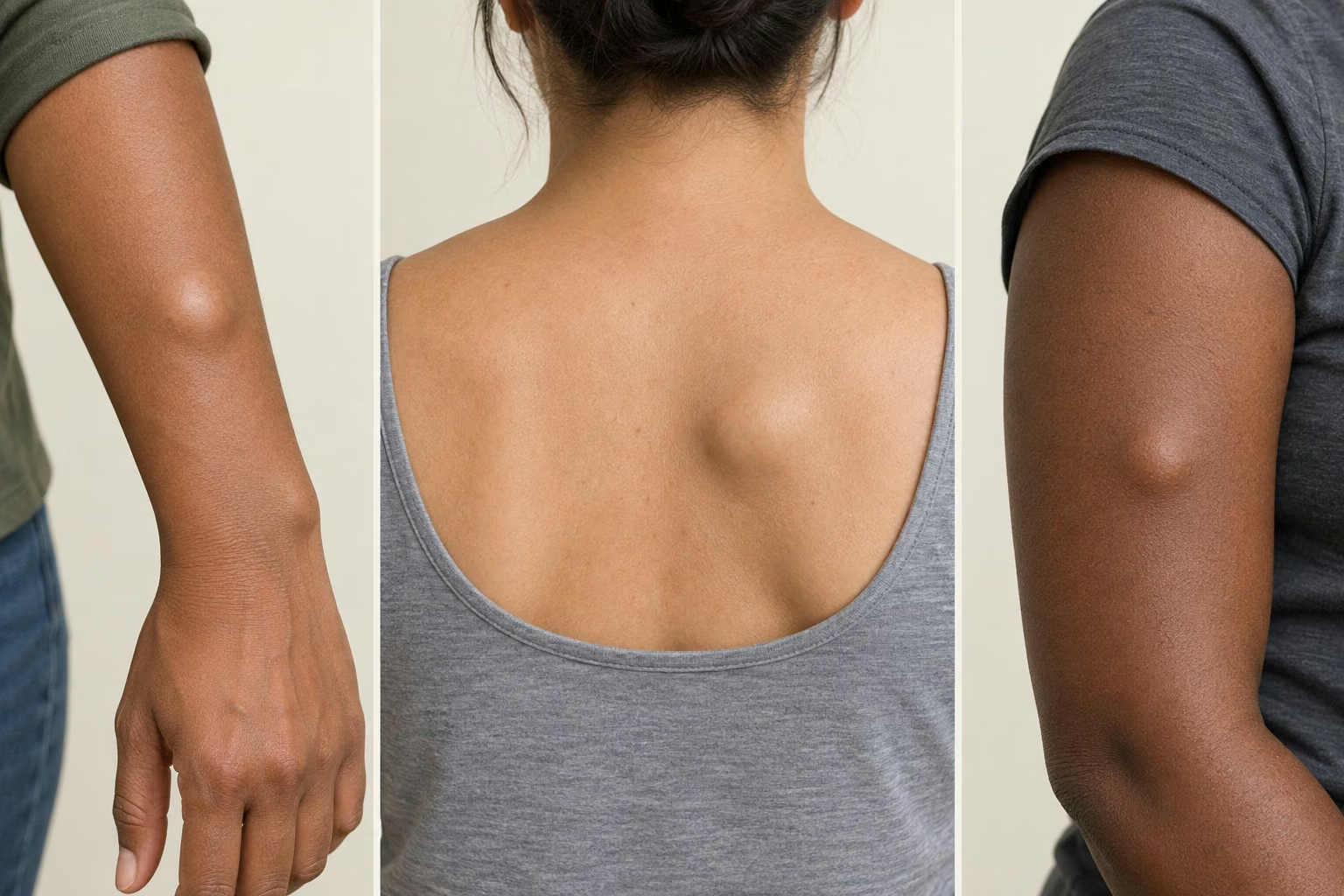
Size, pain, consistency and growth rate matter more than a quick comparison with pictures.
Ultrasound is a common first test; MRI or tissue analysis may be needed when uncertainty remains.
| Feature | Typical lipoma | Needs clarification |
|---|---|---|
| Consistency | Soft or compressible | Very firm or heterogeneous |
| Movement | Moves under the skin | Fixed or deep |
| Evolution | Slow and stable | Rapid growth or return |
| Symptoms | Usually painless | Persistent pain, deficit or limitation |
OPTIONS
When the assessment is typical and there are no symptoms, monitoring size and symptoms may be enough. Agree on what should prompt review.
Removal takes out the mass and allows planned closure. Pain, friction, growth, limitation or cosmetic preference may influence the decision.
The tissue may be sent to a laboratory. The report is part of confirmation and should not be replaced by a visual assumption.
Studies of minimally invasive techniques are series or comparisons in selected groups. They do not mean one technique is best for everyone or promise no scar or recurrence.
EDUCATIONAL IMAGES
READING THE EVIDENCE
Systematic review · diagnostic accuracy
A 2017 systematic review included six studies and reported specificity around 96%. This supports ultrasound for superficial lumps; it does not make every scan conclusive. [2]
Case series · selected patients
A 2015 series treated 48 lipomas in 25 people using liposuction plus excision. Long-term respondents reported no recurrence. The small, uncontrolled series cannot establish superiority over standard excision. [5]
Retrospective cohort · different tumour groups
A study of 141 intramuscular lipomas and 35 atypical lipomatous tumours found different recurrence patterns. Deep lesions and simple superficial lumps should not share one blanket prognosis. [6]
PLAN YOUR CARE
Explain what bothers you most: pain, uncertainty, friction or appearance. Ask which benefit the proposed treatment is expected to deliver and what observation would involve.
Ask for written wound-care instructions, a review date and a contact route for problems. Confirm whether the specimen was sent for analysis and how you will receive the result.
Protect the wound and follow your team’s instructions for washing, dressing and activity. Worsening pain, spreading redness, pus, fever or persistent bleeding need prompt advice.[8]
COMMON QUESTIONS
No question matches that search.
Usually not. A lipoma is a benign tumour made of mature fat cells. A new, deep, firm or rapidly enlarging lump still needs an examination because appearance alone cannot exclude other diagnoses. [7]
No. An epidermoid cyst, hernia, lymph node, vascular lesion and other masses can look similar. Examination guides whether ultrasound or MRI is appropriate. [4]
It is often soft or compressible, mobile over deeper tissues, painless and slow-growing. These features support the diagnosis but do not replace an assessment. [1]
Most do not. Pain can result from pressure on a nerve, friction, local inflammation or a different type of mass. New or progressive pain deserves review. [10]
Yes. The shoulders, trunk, arms, thighs and neck are common sites. Deep or intramuscular lipomas require a different assessment. [10]
Not always. If the diagnosis is secure and it causes no problem, observation is reasonable. Removal may be considered for pain, growth, functional limitation, friction or cosmetic concern. [1]
There is no proven cream or massage that removes a lipoma. Do not puncture it or inject substances. [11]
Ultrasound is useful for small accessible masses. A systematic review found good specificity, but accuracy varies; an indeterminate result needs clinical correlation or another test. [2]
MRI is more useful for deep, large, fixed, painful or atypical lesions and for planning masses near muscle, nerves or vessels. [3]
No. Size alone does not diagnose malignancy, but a large, deep or growing mass deserves organised investigation. [4]
It is a fat tumour with microscopic changes that can behave more locally aggressively than a simple lipoma. Imaging and tissue analysis may be needed to distinguish them. [7]
Many superficial lipomas can be removed under local anaesthesia in an appropriate setting. The plan depends on size, site, depth and health. [11]
Scar size depends on the lesion, location, skin tension and technique. Every removal leaves a mark; a consultation should set realistic expectations. [11]
Often, especially when there is diagnostic uncertainty, greater size, depth or atypical features. Pathology confirms the diagnosis. [7]
A completely removed simple lipoma rarely returns in the same site. Another lipoma can appear elsewhere, and deep or atypical lesions need the follow-up advised by the team. [6]
A painless lipoma usually does not prevent activity. Friction, pain, movement limitation or recent surgery may require temporary adjustment.
Follow the dressing and medication instructions, keep the wound protected, avoid tension on stitches and attend the planned review. Contact the team for worsening pain, spreading redness, pus, fever or persistent bleeding. [8]
No. Cutting, squeezing, tying it off or applying products can cause infection, bleeding, scarring and delayed diagnosis.
Some people have a family tendency or multiple lipomas, but most are isolated. Family history helps guide the assessment. [10]
Seek prompt assessment if the lump grows quickly, becomes hard or fixed, seems deep, is several centimetres across, remains painful, returns after removal or comes with general symptoms. [9]
Record when you noticed the lump, changes, pain and other lumps. Bring previous reports and a list of medicines, allergies and operations. Older photos can help explain the history.
Ask the team first. An assessment does not mean surgery the same day. Preparation depends on the procedure and anaesthesia; do not fast or stop medicines on your own.
The team needs the medicine, dose and reason for treatment to assess bleeding risk. Do not stop anticoagulants, aspirin or other prescribed treatment without individual advice.
They are different diagnoses. Liposarcoma is a malignant fatty-tissue tumour; a confirmed lipoma is benign. The priority is correctly identifying a suspicious mass rather than assuming every lipoma is changing into cancer. [7]
Not if the mass has changed. Bring the previous report and describe what is new. Further imaging or investigation depends on the current findings. [9]
No. It has been studied in selected patients with a reliable diagnosis. Size, depth, capsule and the need for tissue analysis influence the choice. [5]
There is no single timeframe. Site, incision size, stitches and your work matter. Ask for specific dates for dressing changes, review, work and exercise.
Not necessarily. Studies involve selected people, techniques and follow-up periods. They inform the discussion but cannot predict your scar, recurrence or recovery precisely.
Agree on a method and review interval with your clinician. Record dates and changes. Avoid repeatedly pressing or squeezing the lump.
Ask about the likely diagnosis, need for imaging, benefits of removal now, expected scar, tissue analysis, how results will be communicated and the review plan.
PUBLISHED RESEARCH
The links below open the published article or institutional guidance. Research results describe groups and techniques; they do not predict an individual result.
Sources checked on 17 September 2026. Journal articles and institutional guidance are identified separately in each entry.
Medical information is educational and does not replace an in-person examination.
Continue reading with information, frequently asked questions and research references.
INFORMATION FOR SKIN HEALTH
A scalp lump, a body cyst and tiny white facial bumps may need different approaches. Learn when to monitor, investigate or discuss removal.